Morphology of Leydig cells in the testes after in vivo MCP-1 treatment.
Por um escritor misterioso
Descrição

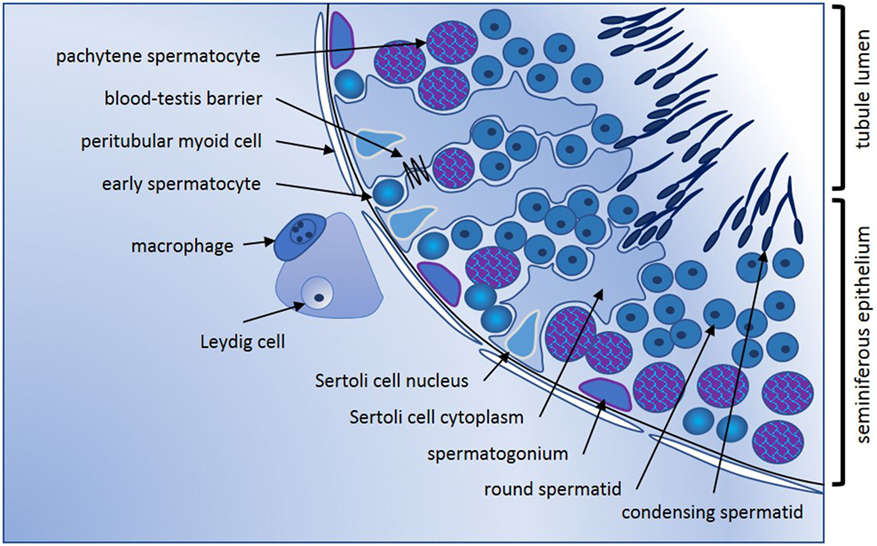
Human testicular peritubular cells: more than meets the eye in: Reproduction Volume 145 Issue 5 (2013)
IJMS, Free Full-Text

Morphology of Leydig cells in the testes after in vivo MCP-1 treatment.

Prenatal exposure to bisphenol AF induced male offspring reproductive dysfunction by triggering testicular innate and adaptive immune responses - ScienceDirect

Effect of TNF on testosterone production. Leydig cells were cultured in
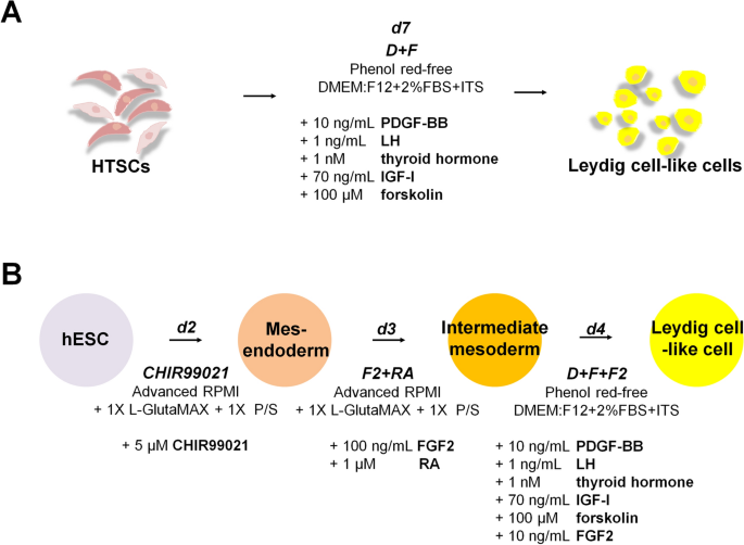
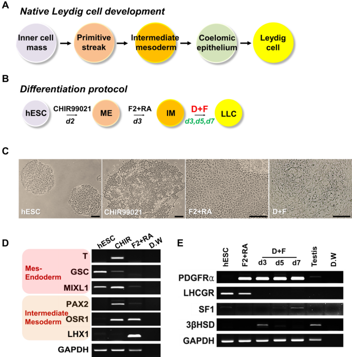
Rapid Differentiation of Human Embryonic Stem Cells into Testosterone-Producing Leydig Cell-Like Cells In vitro

Morphology of Leydig cells in the testes after in vivo MCP-1 treatment.
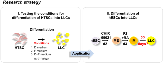
Rapid Differentiation of Human Embryonic Stem Cells into Testosterone-Producing Leydig Cell-Like Cells In vitro

Transcription factor Dmrt1 triggers the SPRY1-NF-κB pathway to maintain testicular immune homeostasis and male fertility
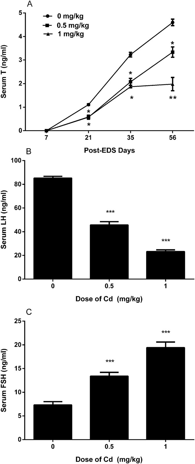
A brief exposure to cadmium impairs Leydig cell regeneration in the adult rat testis
Frontiers Cytokines in Male Fertility and Reproductive Pathologies: Immunoregulation and Beyond
Rapid Differentiation of Human Embryonic Stem Cells into Testosterone-Producing Leydig Cell-Like Cells In vitro

Testicular macrophages are recruited during a narrow time window by fetal Sertoli cells to promote organ-specific developmental functions

Morphology of Leydig cells in the testes after in vivo PTHrP
de
por adulto (o preço varia de acordo com o tamanho do grupo)